پرونده:Schematic diagram of the human eye.png
Schematic_diagram_of_the_human_eye.png (۶۰۰ × ۵۵۰ پیکسل، اندازهٔ پرونده: ۵۴ کیلوبایت، نوع MIME پرونده: image/png)
این پرونده در ویکیانبار موجود است. محتویات صفحهٔ توصیف آن در زیر نمایش داده میشود. |
خلاصه
| توضیح |
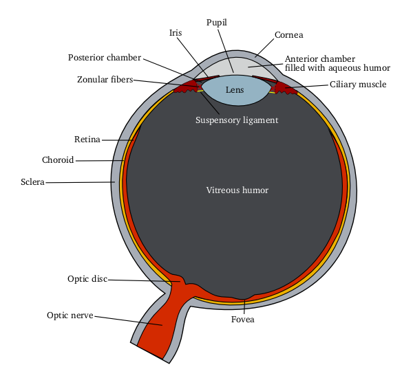
English: Schematic diagram of the human eye
|
|||||
| منبع |
Own work using: |
|||||
| پدیدآور | Delta G | |||||
| دیگر نسخهها |
[] All eye
By languages
For translate
Anterior segment
|


اجازهنامه
| این پرونده با اجازهنامهٔ کریتیو کامانز Attribution-Share Alike 3.0 سازگار نشده منتشر شده است. به تکذیبنامهها رجوع شود. | ||
| ||
| این برچسب مجوز بهعنوان بخشی از روزآمدسازی مجوز GFDL، به این پرونده افزوده شد. |

|
اجازهٔ کپی، پخش و/یا تغییر این سند تحت شرایط مجوز مستندات آزاد گنو، نسخهٔ ۱٫۲ یا هر نسخهٔ بعدتری که توسط بنیاد نرمافزار آزاد منتشر شده؛ بدون بخشهای ناوردا (نامتغیر)، متون روی جلد، و متون پشت جلد، اعطا میشود. یک کپی از مجوز در بخشی تحت عنوان مجوز مستندات آزاد گنو ضمیمه شده است. به تکذیبنامهها رجوع شود. |
Eye Anatomy
A guide to the many parts of the human eye and how they function.
The ability to see is dependent on the actions of several structures in and around the eyeball. The graphic below lists many of the essential components of the eye's optical system.
When you look at an object, light rays are reflected from the object to the cornea, which is where the miracle begins. The light rays are bent, refracted and focused by the cornea, lens, and vitreous. The lens' job is to make sure the rays come to a sharp focus on the retina. The resulting image on the retina is upside-down. Here at the retina, the light rays are converted to electrical impulses which are then transmitted through the optic nerve, to the brain, where the image is translated and perceived in an upright position!
Think of the eye as a camera. A camera needs a lens and a film to produce an image. In the same way, the eyeball needs a lens (cornea, crystalline lens, vitreous) to refract, or focus the light and a film (retina) on which to focus the rays. If any one or more of these components is not functioning correctly, the result is a poor picture. The retina represents the film in our camera. It captures the image and sends it to the brain to be developed. The macula is the highly sensitive area of the retina. The macula is responsible for our critical focusing vision. It is the part of the retina most used. We use our macula to read or to stare intently at an object.
source: http://www.stlukeseye.com/Anatomy.asp
تاریخچهٔ پرونده
روی تاریخ/زمانها کلیک کنید تا نسخهٔ مربوط به آن هنگام را ببینید.
| تاریخ/زمان | بندانگشتی | ابعاد | کاربر | توضیح | |
|---|---|---|---|---|---|
| کنونی | ۵ ژوئن ۲۰۰۶، ساعت ۱۲:۰۰ | | ۶۰۰ در ۵۵۰ (۵۴ کیلوبایت) | Eliashc | optimized using optipng. |
| ۱۴ مارس ۲۰۰۵، ساعت ۰۵:۳۷ |  | ۶۰۰ در ۵۵۰ (۷۳ کیلوبایت) | Delta G | Schematic diagram of the human eye(zonule fibers -> zonular fibers(more common)) | |
| ۱۴ مارس ۲۰۰۵، ساعت ۰۴:۵۸ |  | ۶۰۰ در ۵۵۰ (۷۲ کیلوبایت) | Delta G | Schematic diagram of the human eye(smaller, vitreous fluid -> vitreous humor) | |
| ۱۴ مارس ۲۰۰۵، ساعت ۰۴:۳۶ |  | ۸۶۶ در ۷۹۳ (۱۱۵ کیلوبایت) | Delta G | Schematic diagram of the human eye |
کاربرد پرونده
صفحهٔ زیر از این تصویر استفاده میکند:
کاربرد سراسری پرونده
ویکیهای دیگر زیر از این پرونده استفاده میکنند:
- کاربرد در ar.wikipedia.org
- کاربرد در en.wikipedia.org