پرونده:Electron micrograph of neuromuscular junction (cross-section).jpg
تفکیکپذیری بالاتری در دسترس نیست.
Electron_micrograph_of_neuromuscular_junction_(cross-section).jpg (۴۳۳ × ۲۸۹ پیکسل، اندازهٔ پرونده: ۹۵ کیلوبایت، نوع MIME پرونده: image/jpeg)
این پرونده در ویکیانبار موجود است. محتویات صفحهٔ توصیف آن در زیر نمایش داده میشود. |
.jpg?uselang=fa)
خلاصه
| توضیح |
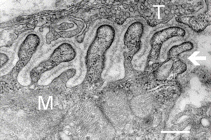
English: Electron micrograph showing a cross-section through the neuromuscular junction. T is the axon terminal, M is the muscle fiber. The arrow shows junctional folds with basal lamina. Postsynaptic densities are visible on the tips between the folds. The scale is 0.3 µm. |
| تاریخ | Originally uploaded to en.wikipedia on ۱۰ مارس ۲۰۰۶. |
| منبع | Synapse Web at the National Institute of Mental Health, National Institutes of Health; originally from en.wikipedia; description page is/was here. |
| پدیدآور | National Institute of Mental Health; originally uploaded by Nrets at en.wikipedia. |
اجازهنامه
This image is a work of the National Institutes of Health, part of the United States Department of Health and Human Services, taken or made as part of an employee's official duties. As a work of the U.S. federal government, the image is in the public domain.
 |
||
| این پرونده تحت قانون حق تکثیر محدودیت آزاد منتشر شده که شامل تمامی حقوق مربوطه و حقوق نزدیک به آن میشود. | ||
سیاهه بارگذاری اصلی
(All user names refer to en.wikipedia)
- 2006-03-10 20:07 Nrets 433×289×8 (97758 bytes) Electron micrograph showing a cross section through the neuromuscular junction. T is the axon terminal, M is the muscle fiber. The arrow shows junctional folds with basal lamina. Postsynaptic densities are visible on the tips between the folds. Scale is 0
تاریخچهٔ پرونده
روی تاریخ/زمانها کلیک کنید تا نسخهٔ مربوط به آن هنگام را ببینید.
| تاریخ/زمان | بندانگشتی | ابعاد | کاربر | توضیح | |
|---|---|---|---|---|---|
| کنونی | ۲۲ مارس ۲۰۰۷، ساعت ۰۳:۴۱ | | ۴۳۳ در ۲۸۹ (۹۵ کیلوبایت) | Fran Rogers | {{Information |Description=Electron micrograph showing a cross section through the neuromuscular junction. T is the axon terminal, M is the muscle fiber. The arrow shows junctional folds with basal lamina. Postsynaptic densities are visible on the tips be |
کاربرد پرونده
صفحهٔ زیر از این تصویر استفاده میکند:
کاربرد سراسری پرونده
ویکیهای دیگر زیر از این پرونده استفاده میکنند:
- کاربرد در ar.wikipedia.org
- کاربرد در cs.wikipedia.org
- کاربرد در de.wikipedia.org
- کاربرد در en.wikipedia.org
- کاربرد در es.wikipedia.org
- کاربرد در gl.wikipedia.org
- کاربرد در he.wikipedia.org
- کاربرد در ko.wikipedia.org
- کاربرد در pt.wikipedia.org
- کاربرد در ru.wikipedia.org
- کاربرد در uk.wikipedia.org
- کاربرد در zh.wikipedia.org
.jpg)