پرونده:Gray491.png
Gray491.png (۵۰۰ × ۴۳۸ پیکسل، اندازهٔ پرونده: ۶۳ کیلوبایت، نوع MIME پرونده: image/png)
این پرونده در ویکیانبار موجود است. محتویات صفحهٔ توصیف آن در زیر نمایش داده میشود. |
خلاصه
| توضیح |
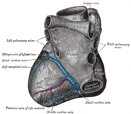
Deutsch: Blick von hinten auf das Herz. Darstellung von Henry Gray. |
||||||||||||||||||||
| Plate | 491 | ||||||||||||||||||||
| تاریخ | پیش از ۱۸۵۸ | ||||||||||||||||||||
| منبع |
|
||||||||||||||||||||
| پدیدآور |
|
||||||||||||||||||||
.jpg)
کتاب
| هنری گری: Gray's Anatomy (20th edition)
|
|||||||||||||||||||||||
|---|---|---|---|---|---|---|---|---|---|---|---|---|---|---|---|---|---|---|---|---|---|---|---|
| پدیدآور |
|
-_Title_page.png) | |||||||||||||||||||||
| ویرایشگر |
Revised by Warren H. Lewis |
||||||||||||||||||||||
| تصویرگر |
|
||||||||||||||||||||||
| عنوان | |||||||||||||||||||||||
| نسخه |
20 |
||||||||||||||||||||||
| ناشر | |||||||||||||||||||||||
| نوع شیء |
ویراست |
||||||||||||||||||||||
| بازنگری صفحه | list of all the plates | ||||||||||||||||||||||
| زبان |
زبان انگلیسی |
||||||||||||||||||||||
| تاریخ انتشار |
۱۹۱۸ |
||||||||||||||||||||||
| محل انتشار |
فیلادلفیا / نیویورک |
||||||||||||||||||||||
| منبع | Bartleby | ||||||||||||||||||||||

اجازهنامه
This image is in the public domain because it is a mere mechanical scan or photocopy of a public domain original, or – from the available evidence – is so similar to such a scan or photocopy that no copyright protection can be expected to arise. The original itself is in the public domain for the following reason:
This tag is designed for use where there may be a need to assert that any enhancements (eg brightness, contrast, colour-matching, sharpening) are in themselves insufficiently creative to generate a new copyright. It can be used where it is unknown whether any enhancements have been made, as well as when the enhancements are clear but insufficient. For known raw unenhanced scans you can use an appropriate {{PD-old}} tag instead. For usage, see Commons:When to use the PD-scan tag.  | ||||
The coronary sinus is a collection of veins joined together to form a large vessel that collects blood from the myocardium of the heart. It is present in humans and other animals. It delivers deoxygenated blood to the Right atrium in conjunction with the superior and inferior vena cava.
The coronary sinus opens into the right atrium, between the inferior vena cava and the atrio-ventricular orifice. It returns the blood from the substance of the heart, and is protected by a semicircular fold of the lining membrane of the auricle, the coronary valve (the valve of Thebesius). The sinus, before entering the auricle, is considerably dilated - nearly to the size of the end of the little finger. Its wall is partly muscular, and at its junction with the great coronary vein is somewhat constricted and furnished with a valve consisting of two unequal segments.(Gray 462)
Location: It is located in the right atrium and runs transversely in the groove between the left atrium and ventricle on the posterior surface of the heart.
The coronary sinus orifice (opening) is just superior to the septal leaflet of the tricuspid valve. The coronary sinus orifice is also known as the ostium of the coronary sinus, and is guarded by the Thebesian valve.
Drainage: It receives blood mainly from the small, middle, great and oblique cardiac veins. It also receives blood from the left marginal vein and the left posterior ventricular vein. The anterior cardiac veins drain directly into the right atrium. (Some small veins drain into any of the four chambers of the heart.)
It drains into the right atrium on the posterior, inferior surface, medial to the inferior vena cava opening.
تاریخچهٔ پرونده
روی تاریخ/زمانها کلیک کنید تا نسخهٔ مربوط به آن هنگام را ببینید.
| تاریخ/زمان | بندانگشتی | ابعاد | کاربر | توضیح | |
|---|---|---|---|---|---|
| کنونی | ۲۳ ژانویهٔ ۲۰۰۷، ساعت ۲۰:۳۵ | | ۵۰۰ در ۴۳۸ (۶۳ کیلوبایت) | Pngbot | optimized with optipng |
| ۱۱ فوریهٔ ۲۰۰۶، ساعت ۰۶:۲۶ |  | ۵۰۰ در ۴۳۸ (۱۰۰ کیلوبایت) | Arcadian | {{Gray's Anatomy plate}} |
کاربرد پرونده
صفحههای زیر از این تصویر استفاده میکنند:
کاربرد سراسری پرونده
ویکیهای دیگر زیر از این پرونده استفاده میکنند:
- کاربرد در ar.wikipedia.org
- کاربرد در bg.wikipedia.org
- کاربرد در bn.wikipedia.org
- کاربرد در bs.wikipedia.org
- کاربرد در cv.wikipedia.org
- کاربرد در de.wikibooks.org
- کاربرد در el.wikipedia.org
- کاربرد در en.wikipedia.org
- Coronary circulation
- Coronary sinus
- Oblique vein of the left atrium
- Posterior descending artery
- Circumflex branch of left coronary artery
- Vital heat
- Posterior interventricular sulcus
- Left marginal artery
- Smallest cardiac veins
- Vascular remodelling in the embryo
- Crux cordis
- User:Bob K31416/BH
- User:Walkerc84/sandbox
- User:Was a bee/Gray
- کاربرد در es.wikipedia.org
- کاربرد در it.wikipedia.org
- کاربرد در ja.wikipedia.org
- کاربرد در ko.wikipedia.org
- کاربرد در nl.wikipedia.org
- کاربرد در nn.wikipedia.org
- کاربرد در pl.wikipedia.org
- کاربرد در pt.wikipedia.org
- کاربرد در ro.wikipedia.org
نمایش استفادههای سراسری از این پرونده.